Muscles Soleus. Anatomy & Physiology
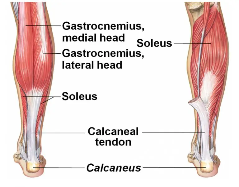
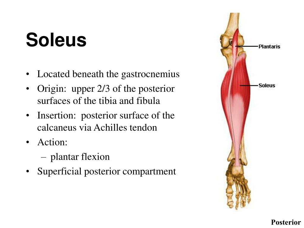
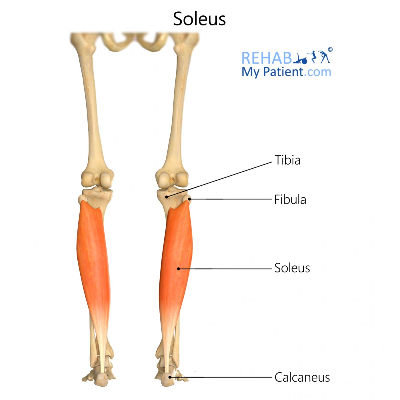
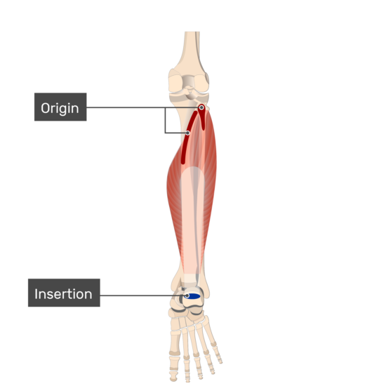
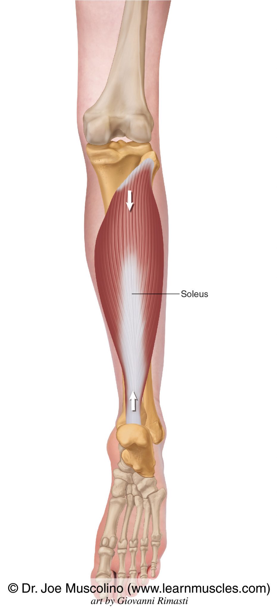
Origin. The soleus muscle originates from the head and upper third of the posterior border of the fibula, soleal line and middle third of the medial border of the tibia and tendinous arch between the fibula and tibia. Insertion. The soleus joins with the gastrocnemius, and both muscles form a common tendon called the calcaneal or Achilles.
5 Great Exercises to Strengthen the Soleus What, Why, and How
The soleus is a large muscle on the back of your lower leg. This powerful muscle arises from the back of your shin bone and attaches to your heel bone as part of the Achilles tendon. The soleus muscle is active during activities like walking, running, and jumping. Injury to the soleus may affect your ability to perform these basic but necessary.

Soleus Muscle Anatomy Origin, Insertion, Action The Wellness Digest
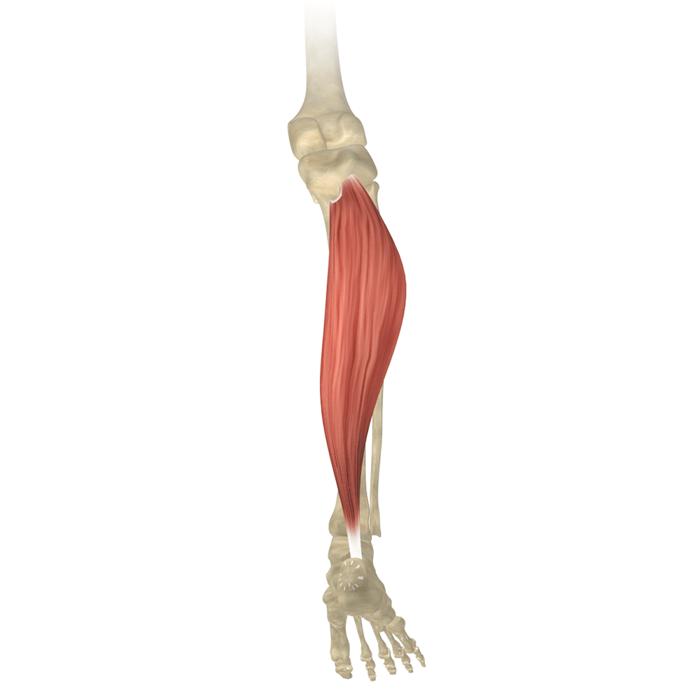
The soleus muscle is a wide flat leg muscle found on the posterior leg.. It runs from just below the knee to the heel and lays immediately deep to the gastrocnemius.These two muscles, along with the plantaris muscle, belong to the group of superficial posterior compartment calf muscles. Soleus' contraction results in strong plantar flexion.It also allows us to maintain an upright posture due.
PPT MUSCLES OF THE ANKLE AND FOOT PowerPoint Presentation, free download ID152691
Soleus Proximal Origin. The soleus muscle has two proximal origins, one fibular and the other tibial. The fibular origin, which is higher than the tibial one, originates through a thick tendon band in the posterior sector of the fibular head, and through aponeurotic fibers from the lateral border of the fibula and the posterior intermuscular.
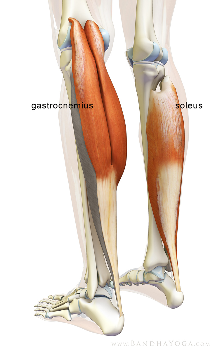
Image Gallery soleus muscle
The soleus is a complex, multi-pennate muscle in humans, usually having a separate (posterior) aponeurosis from the gastrocnemius muscle. Most soleus muscle fibers originate from each side of the anterior aponeurosis, attached to the tibia and fibula. [1] [2] Other fibers originate from the posterior (back) surfaces of the head of the fibula.
Soleus Muscle Attachments, Actions & Innervation
Origin and Insertion: The soleus muscle is a flat, broad muscle that forms the deep layer of the posterior calf. Its origin lies on the posterior surfaces of the tibia and fibula, specifically along the proximal two-thirds of the fibula and the posterior border of the tibia. This origin site is commonly referred to as the "soleal line" on the.

Soleus Anatomy Origin, Insertion, Action, Innervation The Wellness Digest
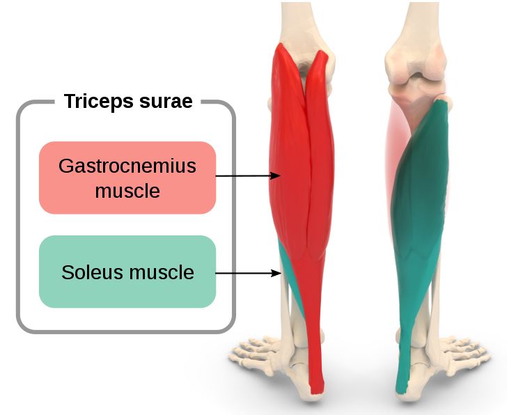
Located in superficial posterior compartment of the leg Soleus is a powerful lower limb muscle, which is situated deep to the gastronemius muscle. Together with gastronemius and plantaris, it forms the calf muscle or triceps surae. It runs from back of the knee to the ankle and is multipennate. The soleus has the greatest physiological cross.
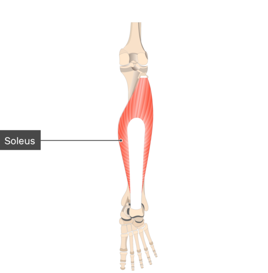
Soleus Rehab My Patient
⭐ Soleus Muscle Anatomy ⭐💪Origin: Head of fibula, posterior surface of fibula, soleal line & medial border of tibia💪Insertion: Posterior surface of the cal.

Image Gallery soleus origin
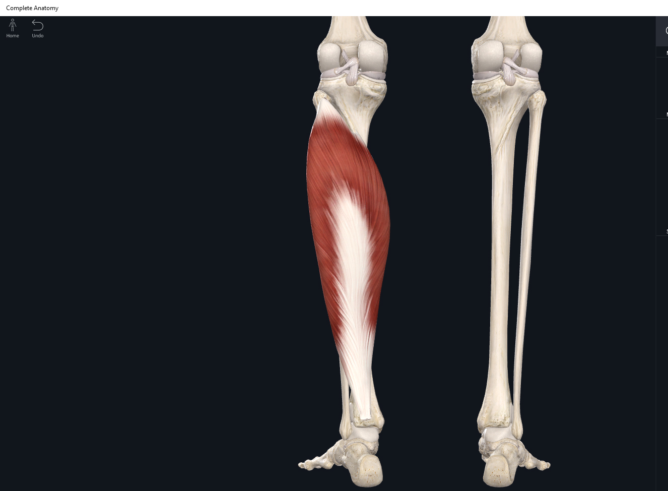
Origin of Soleus Muscle. Description. Posterior aspect of proximal end of fibula. Complete Anatomy. The world's most advanced 3D anatomy platform. Soleus Muscle . Calcaneal Tendon . Complete Anatomy. The world's most advanced 3D anatomy platform. Try it for Free. Useful links. Submit your paper (opens in new tab/window) Shop Books & Journals.
Soleus Muscle Attachments, Actions & Innervation
The soleus is a muscle within the superficial compartment of the posterior leg.. It is a flat muscle located underneath the gastrocnemius, and gets its name from its resemblance to a sole - a flat fish. Attachments: Originates from the soleal line of the tibia and proximal fibula.The muscle converges with the fibres of the gastrocnemius to form the calcaneal tendon, which inserts onto the.
illnesshacker Learn More, Be Sure!!!.
The soleus muscle is one of the calf muscles (triceps surae) in the superficial posterior compartment of the leg which sits deep to gastrocnemius.It is much bigger than gastrocnemius and is the primary plantar flexor. Summary. origin:. fibular origin: head of fibula and upper third of posterior fibula; tibial origin: medial border of the tibia and posterior border of tibia (soleal line)
Functional anatomy guide Soleus muscle • Bodybuilding Wizard
The soleus muscle also originates from a fibrous band that extends from its origin sites on the fibula and tibia, known as the tendinous arch of soleus. Insertion The fibers of the soleus muscle travel inferiorly and converge with the fibers of the medial and lateral heads of the gastrocnemius muscle to form the calcaneal tendon, which inserts.

The Soleus Muscles, Its Attachments and Actions Yoganatomy
Soleus. Posterior aspect of fibular head, upper 1/4 of posterior surface of fibula, middle 1/3 of medial border of tibial shaft, and from posterior surface of a tendinous arch spanning the two sites of bone origin. Eventually unites with the gastrocnemius aponeurosis to form the Achilles tendon, inserting on the middle 1/3 of the posterior.

Soleus Anatomy Origin, Insertion & Action YouTube
When to see a doctor. The soleus is a muscle in the calf that runs from directly behind the knee to just above the muscles around the ankle. An injury or strain to this muscle can cause pain and.
Soleus Learn Muscles
The muscles were harvested from the right legs of fresh-frozen cadavers of a 67-year old male (muscle 1; muscle length: 36.5 cm) and a 90-year old male (muscle 2; muscle length: 32 cm). After gross dissection, the soleus muscles were fixed by submerging the tissue in a 10% neutral buffered formalin solution for 11 weeks.

Soleus Attachments and Actions 3D Muscle Lab
Last modified: 20 November 2022. The soleus is one of the muscles within the superficial group within the posterior compartment of the leg. The soleus muscle is a large flat muscle which lies immediately deep to the gastrocnemius muscle. The leg is comprised of anterior, lateral and posterior compartments. The posterior (flexor) compartment.
